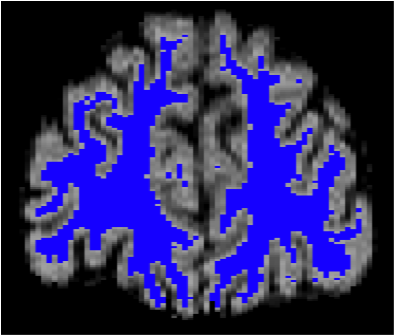
Figure 2. Cross-Section of the human brain showing brain cells (gray matter) in the outer ‘gray’ rim, and brain connections (white matter) in inner ‘blue’ area.
(Credit: Foundas Brain Lab).
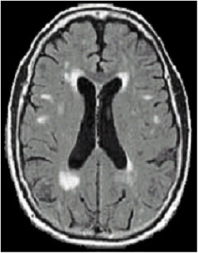
Figure 3. Horizontal brain image showing white matter disease visualized as the punctate ‘white’ areas. These lesions are consistent with white matter hyperintensities seen on MRI brain scans.